Limfoma Non-Hodgkin
Pendahuluan dan Fakta
Limfoma adalah sekumpulan keganasan primer pada kelenjar getah bening dan jaringan limfoid. Berdasarkan tipe histologiknya, limfoma dapat dibagi menjadi dua kelompok besar, yaitu Limfoma Non Hodgkin dan Hodgkin. Pada protokol ini hanya akan dibatasi pada limfoma non-Hodgkin.
Limfoma Non Hodgkin (LNH) merupakan sekumpulan besar keganasan primer kelenjar getah bening dan jaringan limfoid ekstra nodal, yang dapat berasal dari limfosit B, limfosit T, dan sel NK *”natural killer”. Saat ini terdapat 36 entitas penyakit yang dikategorikan sebagai LNH dalam klasifikasi WHO. Di Indonesia, LNH bersama-sama dengan limfoma Hodgkin dan leukemia menduduki urutan peringkat keganasan ke-6.
Patofisiologi
LNH merupakan keganasan primer limfosit yang berasal dari limfosit B, limfosit T dan kadang-kadang berasal dari sel natural killer yang berada dalam sistem limfe dengan gambaran yang sangat heterogen baik secara histologis, gejala, perjalanan klinis, respon terhadap pengobatan maupun prognosis. Seperti keganasan lain, LNH merupakan hasil dari akumulasi kelainan genetika yang bertahap sehingga terjadi pertumbuhan yang tidak terkendali klon sel-sel ganas. Translokasi berulang yang terjadi pada beberapa tingkat deferensiasi sel B merupakan awal dari transformasi maligna. Translokasi ini mengakibatkan deregulasi ekspresi onkogen yang mengontrol proliferasi, survival dan deferensiasi sel. Menariknya, translokasi saja tidak cukup untuk menyebabkan terjadinya limfoma, sehingga diperlukan gangguan genetika sekunder berikutnya untuk terjadinya transformasi maligna seutuhnya.
Gejala Klinis dan Komplikasi
Gejala yang sering ditemukan pada penderita limfoma pada umumnya non-spesifik, diantaranya:
- Penurunan berat badan >10% dalam 6 bulan.
- Demam 38 derajat C >1 minggu tanpa sebab yang jelas.
- Keringat malam banyak.
- Cepat lelah.
- Penurunan nafsu makan.
- Pembesaran kelenjar getah bening yang terlibat.
- Dapat pula ditemukan adanya benjolan yang tidak nyeri di leher, ketiak atau pangkal paha (terutama bila berukuran di atas 2 cm); atau sesak napas akibat pembesaran kelenjar getah bening mediastinum maupun splenomegali.
Tiga gejala pertama harus diwaspadai karena terkait dengan prognosis yang kurang baik, begitu pula bila terdapatnya Bulky Disease (KGB berukuran > 6-10 cm atau mediastinum >33% rongga toraks). Menurut Lymphoma International Prognostic Index, temuan klinis yang mempengaruhi prognosis penderita LNH adalah usia >60 tahun, keterlibatan kedua sisi diafragma atau organ ekstra nodal (Ann Arbor III/IV) dan multifokalitas (>4 lokasi).
Diagnosis
Diagnosis ditegakkan berdasarkan anamnesis, pemeriksaan fisik, pemeriksaan laboratorik, dan Patologi Anatomik.
Pemeriksaan:
1. Anamnesis Umum:
- Pembersaran kelenjar getah bening (KGB) atau organ.
- Malaise umum.
- Berat badan menurun >10% dalam waktu 3 bulan.
- Demam tinggi 38°C selama 1 minggu tanpa sebab.
- Keringat malam.
- Keluhan anemia (lemas, pusing, jantung berdebar)?
- Penggunaan obat-obatan tertentu?
- Khusus:
o Penyakit autoimun (SLE, Sjorgen, Rheumatoid)
o Kelainan darah
o Penyakit infeksi (Toxoplasma, Mononukleosis,Tuberkulosis, Lues, dsb)
o Keadaan defisiensi imun
2. Pemeriksaan Fisik
- Pembesaran KGB.
- Kelainan/pembesaran organ (hati/limpa).
- Performance status: ECOG atau WHO/Karnofsky.
3. Pemeriksaan Diagnostik
A. Biopsi eksisional atau core biopsy:
Biopsi KGB dilakukan cukup pada 1 kelenjar yang paling representatif, superfisial, dan perifer. Jika terdapat kelenjarsuperfisial/perifer yang paling representatif, maka tidak perlu biopsi intraabdominal atau intratorakal. Kelenjar getah bening yang disarankan adalah dari leher dan supraclavicular, pilihan kedua adalah aksila dan pilihan terakhir adalah inguinal.Spesimen kelenjar diperiksa:
a. Rutin, Histopatologi: sesuai klasifikasi WHO terbaru
b. Khusus, Immunohistokimia dan Molekuler (hibridisasi insitu) EBV
Diagnosis awal harus ditegakkan berdasarkan histopatologi dan tidak cukup hanya dengan sitologi. Pada kondisi tertentu dimana KGB sulit dibiopsi, maka kombinasi core biopsy FNAB bersama-sama dengan teknik lain (IHK, Flowcytometri `dan lain-lain) mungkin dapat mencukupi untuk diagnosis.
B. Laboratorium
1. Rutin Hematologi:
o Darah Perifer Lengkap (DPL) : Hb, Ht, leukosit,trombosit, LED, hitung jenis
o Gambaran Darah Tepi (GDT) : morfologi sel darah
o Analisis urin : urin lengkap
2. Kimia klinik:
o SGOT, SGPT, Bilirubin (total/direk/indirek), LDH, protein total, albumin-globulin
o Alkali fosfatase, asam urat, ureum, kreatinin o Gula darah sewaktu
o Elektrolit: Na, K, Cl, Ca, P
o HIV, TBC, Hepatitis C (anti HCV, HBsAg) Khusus:
o Gamma GT
o Serum Protein Elektroforesis (SPE) o Imunoelektroforesa (IEP)
o Tes Coomb
o B2 mikroglobulin
C. Aspirasi Sumsum Tulang (BMP) dan biopsi sumsum tulang dari 2 sisi spina illiaca dengan hasil spesimen minimal panjang 1.5 cm, dan disarankan 2 cm.
D. Radiologi. Untuk pemeriksaan rutin/standard dilakukan pemeriksaan CT Scan thorak/abdomen.Bila fasilitas tersedia, dapat dilakukan PET CT Scan.
E. Konsultasi THT. Bila Cincin Waldeyer terkena dilakukan laringoskopi.
F. Cairan tubuh lain (Cairan pleura, cairan asites, cairan liquor serebrospinal). Jika dilakukan pungsi/aspirasi diperiksa sitologi dengan cara cytospin, disamping pemeriksaan rutin lainnya.
G. Konsultasi jantung. Menggunakan echogardiogram untuk melihat fungsi jantung
Tatalaksana dan Perawatan
Pilihan terapi bergantung pada beberapa hal, antara lain: tipe limfoma (jenis histologi), stadium, sifat tumor (indolen/agresif), usia, dan keadaan umum pasien.
I. LNH INDOLEN / Low grade: (Ki-67 < 30%) Yang termasuk dalam kelompok ini adalah:
- SLL/small lymphocytic lymphoma/CLL = chronic lymphocytic lymphoma.
- MZL (marginal zone lymphoma), nodal, ekstranodal dan splenic).
- Lymphoplasmacytic lymphoma.
- Follicular lymphoma.
- Mycosis Fungoides.
- Primary cutaneous anaplastic large cell lymphoma.
A. LNH INDOLEN STADIUM I DAN II
Radioterapi memperpanjang disease free survival pada beberapa pasien. Standar pilihan terapi :
1. Iradiasi
2. Kemoterapi dilanjutkan dengan radiasi
3. Kemoterapi (terutama pada stadium =2 (menurut kriteria GELF)
4. Kombinasi kemoterapi dan imunoterapi
5. Observasi
B. LNH INDOLEN / low grade STADIUM II bulky, III, IV Standar pilihan terapi :
1. Observasi (kategori 1) bila tidak terdapat indikasi untuk terapi. Termasuk dalam indikasi untuk terapi:
- Terdapat gejala.
- Mengancam fungsi organ.
- Sitopenia sekunder terhadap limfoma.
- Bulky.
- Progresif.
- Uji Klinik.
2. Terapi yang dapat diberikan:
1. Rituximab dapat diberikan sebagai kombinasi terapi lini pertama yaitu R-CVP. Pada kondisi dimana Rituximab tidak dapat diberikan maka kemoterapi kombinasi merupakan pilihan pertama misalnya: COPP, CHOP dan FND.
2. Purine nucleoside analogs (Fludarabin) pada LNH primer
3. Alkylating agent oral (dengan/tanpa steroid), bila kemoterapi kombinasi tidak dapat diberikan/ditoleransi (cyclofosfamid, chlorambucil)
4. Rituximab maintenance dapat dipertimbangkan
5. Kemoterapi intensif ± Total Body irradiation (TBI) diikuti dengan stem cell resque dapat dipertimbangkan pada kasus tertentu
6. Raditerapi paliatif, diberikan pada tumor yang besar (bulky) untuk mengurangi nyeri/obstruksi.
C. LNH INDOLEN/ low grade RELAPS
Standar pilihan terapi:
1. Radiasi paliatif
2. Kemoterapi
3. Transplantasi sumsum tulang
II. LNH AGRESIF / High grade: (Ki-67 > 30%) Yang termasuk dalam kelompok ini adalah:
- MCL (Mantle cell lymphoma, pleomorphic variant).
- Diffuse large B cell lymphoma, Follicular lymphoma gr III, B cell lymphoma unclassifiable with features between diffuse large B cell and Burkitt,
- T cell lymphomas.
A. LNH STADIUM I DAN II
Pada kondisi tumor non bulky (diameter tumor <7.5cm) dengan kriteria: pasien muda risiko rendah atau rendah-menengah (aaIPI score =1) dan risiko tinggi atau menengah-tinggi (aaIPI =2), bila fasilitas memungkinkan, kemoterapi kombinasi R-CHOP 6 siklus merupakan protokol standar saat ini serta dapat dipertimbangkan pemberian radioterapi (untuk konsolidasi), atau kemoterapi 3 siklus dilanjutkan dengan radioterapi.
B. LNH STADIUM I-II (BULKY), III DAN IV
• Bila memungkinkan, pemberian kemoterpi RCHOP 6siklus ± radioterapi konsolidasi, dipertimbangkan pada stadium I dan II
• Uji klinik pada stadium III dan IV
C. LNH REFRAKTER/RELAPS
• Pasien LNH refrakter yang gagal mencapai remisi, dapat diberikan terapi salvage dengan radioterapi jika area yang terkena tidak ekstensif. Terapi pilihan bila memungkinakan adalah kemoterapi salvage diikuti dengan transplantasi sumsum tulang
• Kemoterapi salvage seperti R-DHAP maupun R-ICE
III. LNH “LEUKEMIA-LIKE”: Lymphoblastic, Burkitt, “double hit” lymphoma.
• High dose chemotherapy plus radioterapi diikuti dengan transplantasi sumsum tulang
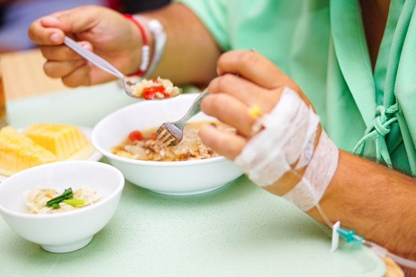
Dukungan Nutrisi. Status gizi merupakan salah satu faktor yang berperan penting pada kualitas hidup pasien kanker.
Rehabilitasi Medik. Rehabilitasi medik bertujuan untuk mengoptimalkan pengembalian gangguan kemampuan fungsi dan aktivitas kehidupan sehari-hari serta meningkatkan kualitas hidup pasien dengan cara aman & efektif, sesuai kemampuan fungsi yang ada. Pendekatan rehabilitasi medik dapat diberikan sedini mungkin sejak sebelum pengobatan definitif diberikan dan dapat dilakukan pada berbagai tahapan & pengobatan penyakit yang disesuaikan dengan tujuan penanganan rehabilitasi kanker: preventif, restorasi, suportif atau paliatif.
Edukasi, yang meliputi:
1. Kemoterapi:
- Efek samping kemoterapi yang mungkin muncul (CPIN, dsb)
- Latihan yang perlu dilakukan untuk menghindari gangguan kekuatan otot (lihat prinsip rehabilitasi medik)
2. Nutrisi . Edukasi jumlah nutrisi , jenis dan cara pemberian nutrisi sesuai dengan kebutuhan
3. Lainnya. Anjuran untuk kontrol rutin pasca pengobatan
Referensi:
1. Komite Penganggulangan Kanker Nasional. Panduan Penatalaksanaan Limfoma Non-Hodgkin. Internet [Cited 24/8/2021]. Available from: http://kanker.kemkes.go.id/guidelines_read.php?id=2&cancer=6
2. Karlina Isabella. Ki-67 Sebagai Parameter Prognosis pada Limfoma Non-Hodgkin. Internet [Cited 24/8/2021]. Available from: https://simdos.unud.ac.id/uploads/file_penelitian_1_dir/ef83468df2f3d04c7a6c61bd0ad56aa4.pdf


