


Trombosis Vena Dalam (DVT)
Pendahuluan dan Fakta
Trombosis vena dalam atau deep vein thrombosis (DVT) terjadi ketika bekuan darah (trombus) terbentuk di satu atau lebih vena dalam di tubuh, seringkali pada tungkai bawah. Trombosis vena dalam dapat menyebabkan nyeri atau bengkak, namun terkadangan tidak timbul gejala yang signifikan.
Insidens DVT di Eropa dan Amerika Serikat kurang lebih 50 per 100.000 populasi/tahun. Angka kejadian DVT meningkat sesuai umur, sekitar 1 per 10.000 – 20.000 populasi pada umur di bawah 15 tahun hingga 1 per 1000 populasi pada usia di atas 70 tahun. Insidens DVT pada ras Asia dan Hispanik dilaporkan lebih rendah dibandingkan pada ras Kaukasia, Afrika-Amerika Latin, dan Asia Pasifik. Tidak ada perbedaan insidens yang signifikan antara pria dan wanita.
Patofisiologi
Trombosis vena biasanya terdiri dari fibrin, sel darah merah, dan beberapa komponen trombosit dan leukosit. Terdapat tiga hal yang berperan dalam proses terjadinya trombosis (Virchow’s Triad):
1. Stasis vena. Aliran darah vena cenderung lambat, bahkan dapat stasis terutama di daerah yang mengalami imobilisasi cukup lama. Stasis vena merupakan faktor predisposisi terjadinya trombosis lokal, karena dapat mengganggu mekanisme pembersihan aktivitas faktor pembekuan darah sehingga memudahkan terbentuknya trombosis.
2. Kerusakan pembuluh darah. Kerusakan pembuluh darah dapat berperan dalam proses pembentukan trombosis vena, melalui:
- Trauma langsung yang mengakibatkan faktor pembekuan
- Aktivasi sel endotel oleh sitokin yang dilepaskan sebagai akibat kerusakan jaringan dan proses peradangan.
3. Perubahan daya beku darah. Dalam keadaan normal terdapat keseimbangan sistem pembekuan darah dan sistem fibrinolisis. Kecenderungan trombosis terjadi apabila aktivitas pembekuan darah meningkat atau aktivitas fibrinolisis menurun. DVT sering terjadi pada kasus aktivitas pembekuan darah meningkat, seperti pada hiperkoagulasi, defisiensi anti-trombin III, defisiensi protein-C, defisiensi protein S, dan kelainan plasminogen.
Etiologi
Apapun itu yang menghalangi mengalirnya darah dengan baik atau membeku dapat menyebabkan penggumpalan darah. Penyebab utama pada DVT adalah kerusakan vena akibat pembedahan dan kerusakan akibat infeksi atau cedera.
Faktor Risiko
Terdapat beberapa faktor yang meningkatkan risiko terjadinya DVT. Beberapa faktornya meliputi:
- Usia
- Kurangnya pergerakan
- Trauma atau pembedahan
- Kehamilan
- Pil kontrasepsi
- Obesitas
- Merokok
- Kanker
- Gagal jantung
- Inflammatory bowel disease
- Riwayat keluarga dengan DVT atau emboli paru
- Genetik
Tanda dan Gejala
Tidak semua orang dengan DVT menunjukkan gejala, namun terdapat beberapa hal yang bisa menjadi perhatian lebih:
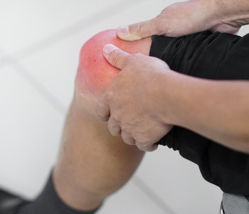
- Pembengkakan pada kaki atau lengan yang muncul secara tiba-tiba
- Nyeri atau rasa pegal saat berdiri atau berjalan
- Rasa hangat pada area yang nyeri
- Pembesaran pembuluh darah
- Kulit yang tampak merah atau kebiruan
Jika bekuan darah terlepas dan bergerak melalui aliran darah, bekuan darah tersebut dapat tersangkut di pembuluh darah paru-paru, yang dikenal dengan emboli paru dan bisa berakibat fatal.
Diagnosis
Penegakkan diagnosis pada DVT diawali dengan anamnesis, pemeriksaan fisik, dan diikuti dengan pemeriksaan penunjang. Anamnesis dilakukan untuk mengetahui gejala penyakit, onset terjadinya penyakit, adanya riwayat trauma atau keluarga dengan DVT, dan masih banyak pertanyaan lainnya. Setelah dilakukan anamnesis, dokter akan melakukan pemeriksaan fisik pada kaki dan area tubuh bagian bawah untuk memeriksa gejalanya. Jika ada pembengkakan, nyeri tekan, atau perubahan warna kulit, kemungkinan besar akan dilakukan beberapa pemeriksaan penunjang untuk membantu diagnosis. Beberapa pemeriksaan penunjang yang dilakukan meliputi:
- Pemeriksaan D-dimer. Pemeriksaan D-dimer dilakukan untuk melihat adanya peningkatan pada D-dimer atau tidak. Peningkatan D-dimer menandakan adanya pembekuan darah.
- Duplex Ultrasound. Pemeriksaan ini merupakan pemeriksaan pencitraan utama untuk mendiagnosis DVT. Ultrasound menggunakan gelombang suara untuk membuat gambar dari bagian dalam tubuh. Gambar-gambar tersebut dapat menunjukkan darah di pembuluh darah dan penggumpalan apa pun.
- MRI. MRI biasanya dilakukan untuk DVT di perut bagian bawah atau daerah perut. Pemeriksaan ini bukanlah standar umum dari pemeriksaan DVT lainnya.
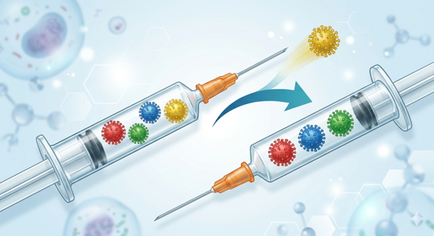
- Venografi. Pemeriksaan ini jarang dilakukan karena sangat invasif. Dokter akan menyuntikkan kontras ke pembuluh darah dan melakukan rontgen untuk melihat apakah kontras mengalir dengan baik melalui pembuluh darah. Hal ini memungkinkan dokter untuk melihat apakah terjadi penggumpalan.
Tata Laksana dan Perawatan
Hanya dilakukan pada kasus yang diagnosisnya sudah jelas ditegakkan mengingat obatobatan dapat menimbulkan efek samping serius. Tujuan tatalaksana DVT fase akut adalah:
1. Menghentikan bertambahnya trombus
2. Membatasi bengkak tungkai yang progresif
3. Melisis dan membuang bekuan darah serta mencegah disfungsi vena atau terjadinya sindrom pasca-trombosis
4. Mencegah terjadinya emboli
a. Non-farmakologis. Penatalaksanaan non-farmakologis terutama ditujukan untuk mengurangi morbiditas pada serangan akut serta mengurangi insidens posttrombosis syndrome yang biasanya ditandai dengan nyeri, kaku, edema, parestesi, eritema, dan edema. Untuk mengurangi keluhan dan gejala trombosis vena pasien dianjurkan untuk istirahat di tempat tidur (bedrest), meninggikan posisi kaki, dan dipasang compression stocking dengan tekanan kira-kira 40 mmHg
b. Farmakologis. Meluasnya proses trombosis dan emboli paru dapat dicegah dengan antikoagulan dan fibrinolitik.
1. Unfractionated Heparin. Terapi unfractionated heparin berdasarkan berat badan dan dosisnya dititrasi berdasarkan nilai Activated Partial Thromboplastin Time (APTT). Nilai APTT yang diinginkan adalah 1,5-2,5 kontrol.
2. Low-Molecular-Weight Heparin (LMWH). Dibandingkan dengan unfractionated heparin, LMWH lebih menguntungkan karena waktu paruh biologis lebih panjang, dapat diberikan subkutan satu atau dua kali sehari, dosisnya pasti dan tidak memerlukan pemantauan laboratorium. Pada pasien DVT, heparin subkutan tidak kurang efektif dibandingkan unfractionated heparin infus kontinyu
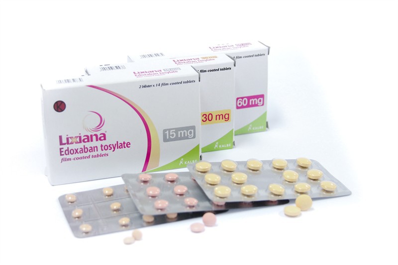
3. Warfarin. Warfarin adalah obat pilihan untuk antikoagulasi akut. Pemberian warfarin segera setelah diagnosis DVT ditegakkan, namun kerjanya memerlukan satu minggu atau lebih. Oleh karena itu, LMWH diberikan bersamaan sebagai terapi penghubung hingga warfarin mencapai dosis terapeutiknya.
4. Terapi Trombolitik. Tidak seperti antikoagulan, obat-obat trombolitik menyebabkan lisisnya trombus secara langsung dengan peningkatan produk plasmin melalui aktivasi plasminogen. Obat-obat trombolitik yang direkomendasikan FDA meliputi streptokinase, recombinant tissue plasminogen activator (rt-PA), dan urokinase
c. Trombektomi. Terapi open surgical thrombectomy direkomendasikan untuk DVT yang memiliki kriteria di antaranya adalah DVT iliofemoral akut, tetapi terdapat kontraindikasi trombolitik atau trombolitik ataupun mechanical thrombectomy gagal, lesi tidak dapat diakses oleh kateter, trombus sukar dipecah dan kontraindikasi antikoagulan.
Referensi:
- Mayo Clinic. Deep vein thrombosis [Internet]. 2022. Available from: https://www.mayoclinic.org/diseases-conditions/deep-vein-thrombosis/symptoms-causes/syc-20352557
- National Health System UK. DVT (deep vein thrombosis) [Internet]. 2023. Available from: https://www.nhs.uk/conditions/deep-vein-thrombosis-dvt/
- Shaban D. Deep vein thrombosis (DVT): Symptoms, causes, treatment. WebMD [Internet]. 2023. Available from: https://www.webmd.com/dvt/what-is-dvt-and-what-causes-it
- Jayanegara AP. Diagnosis dan tatalaksana deep vein thrombosis. CDK 2016:244:43(9):652-7