


Penyakit Perlemakan Hati Non-alkohol
Pendahuluan dan Fakta
Penyakit perlemakan hati non-alkohol (non-alcoholic fatty liver disease/ NAFLD) merupakan kondisi steatosis hepatik, inflamasi, dan kerusakan hepatosit (ballooning degeneration). Di negara Barat dan berbagai negara di Asia, perubahan gaya hidup dan diet meningkatkan prevalensi obesitas dan sindrom metabolik yang sejalan dengan peningkatan insidens NAFLD
Kondisi NAFLD tidak hanya dianggap sebagai penyakit hati primer, tetapi juga merupakan bagian dari sindrom metabolik ataupun kondisi resistensi insulin dan penyakit terkait gaya hidup seperti diabetes, dislipidemia, dan hipertensi. NAFLD telah menjadi masalah dunia dengan prevalensi 10-50% yang juga dipengaruhi etnis hingga jenis kelamin dan diperkirakan bertambah sejalan dengan bertambahnya masalah obesitas dan sindrom metabolik di dunia.
Patofisiologi
Resistensi insulin, stres oksidatif, dan inflamasi dipercaya memainkan peran pada patogenesis dan progresi NAFLD. Hipotesis ‘multi-hit’ (yang dulunya disebut sebagai ‘two-hit’) telah digunakan dalam menjelaskan patogenesis NAFLD. Resistensi insulin menyebabkan meningkatnya asam lemak bebas yang diabsorpsi oleh hati, menghasilkan keadaan steatosis sebagai hit pertama (first hit). Hal tersebut dilanjutkan dengan berbagai interaksi kompleks (multiple second hit) yang melibatkan sel hati, sel stelata, sel adiposa, sel kupfer, mediator-mediator inflamasi, dan reactive oxygen species yang dapat menyebabkan inflamasi (NASH) atau berlanjut sirosis.
Resistensi insulin menginisiasi hit pertama. Keadaan resistensi insulin menyebabkan sel adiposa dan sel otot cenderung mengoksidasi lipid, yang menyebabkan pelepasan asam lemak bebas. Asam lemak lalu diabsorpsi oleh hati, menghasilkan keadaan steatosis. Asam lemak bebas di dalam hati dapat terikat dengan trigliserida atau mengalami oksidasi di mitokondria, peroksisom, atau mikrosom.
Produk-produk hasil oksidasi sifatnya berbahaya dan dapat menyebabkan cedera pada hati yang selanjutnya dapat berlanjut menjadi fibrosis. Peroksidasi lipid dan stres oksidatif meningkatkan produksi hidroksineonenal (HNE) dan malondialdehid (MDA) yang meningkatkan fibrosis hati melalui aktivasi oleh sel stelata yang menyebabkan peningkatan produksi transforming growth factor-beta (TGF-ß).
Mediator-mediator inflamasi berperan pada progresi NAFLD. Faktor transkripsi prionflamasi seperti nuclear factor kappa beta (NF-?ß) sering ditemukan meningkat pada pasien NASH. Adiponektin dan tumor necrosis factoralpha (TNF-a) merupakan dua protein proinflamasi yang berkaitan dengan patogenesis NAFLD. Adiponektin merupakan hormon yang dilepaskan oleh sel adiposa yang menurunkan oksidasi asam lemak dan menghambat glukoneogenesis hepatik. Manusia ataupun tikus menunjukkan level adiponektin yang rendah dan berhubungan dengan peningkatan derajat keparahan inflamasi. Pemecahan adiponektin pada tikus menunjukkan peningkatan signifikan derajat steatosis dan inflamasi. TNF-a merupakan mediator inflamasi yang sebagian besar diproduksi oleh makrofag, serta juga diproduksi oleh sel adiposa dan hepatosit. TNF-a menyebabkan cedera pada hati melalui inhibisi transport elektron mitokondria dan pelepasan reactive oxygen species yang menstimulasi peroksidasi lipid.
Inaktivasi sel Kupfer juga berkaitan pada NAFLD dan penurunan kapasitas regenerasi sel hati. Eliminasi sel Kupfer diasosiasikan dengan peningkatan derajat NASH. Fungsi sel Kupfer terganggu pada situasi peningkatan lemak hati yang mungkin disebabkan karena sinusoid hati yang terlalu ‘penuh’ dan menyebabkan paparan antigen berkepanjangan terhadap sel Kupfer serta penurunan aliran keluar sel Kupfer, yang menyebabkan respons inflamasi yang menetap.
Gejala Klinis dan Komplikasi
Seperti pada penyakit hati kronik lainnya, sebagian besar pasien dengan NAFLD adalah asimtomatik. NAFLD biasa ditemukan secara tidak sengaja pada pemeriksaan laboratorium rutin atau pemeriksaan lanjutan dari keadaan-keadaan lain, seperti hipertensi, diabetes, dan obesitas berat. Peningkatan level ALT atau penemuan bukti-bukti NAFLD secara sonografi dapat pula ditemukan pada pemeriksaan batu empe
Gejala yang mungkin muncul biasanya bersifat tidak spesifik. Kelelahan merupakan yang paling sering dilaporkan dan tidak berkorelasi dengan keparahan lesi histologis. Gejala umum lainnya adalah rasa tidak nyaman pada perut kanan atas yang bersifat samar-samar dan tidak dapat dikategorikan pada rasa nyeri tertentu.
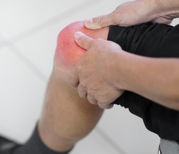
Tidak terdapat tanda patognomonik untuk NASH. Obesitas merupakan abnormalitas yang paling sering ditemukan pada pemeriksaan fisik dan terdapat pada 30-100% pasien. Hepatomegali merupakan hal yang paling sering ditemukan pada pasien dengan gangguan hati. Sebagian kecil pasien juga menunjukkan tanda-tanda stigmata penyakit hati kronik, di mana eritema palmar dan spider nevi adalah yang tersering. Jaundice, asites, asteriksis dan tanda hipertensi portal dapat ditemukan pada pasien dengan sirosis lanjut. Muscle wasting juga dapat ditemukan saat penyakit berlanjut, namun sering tersamar oleh keadaan edema atau obesitas yang telah ada sebelumnya.
Diagnosis
1. Pemeriksaan Laboratorium
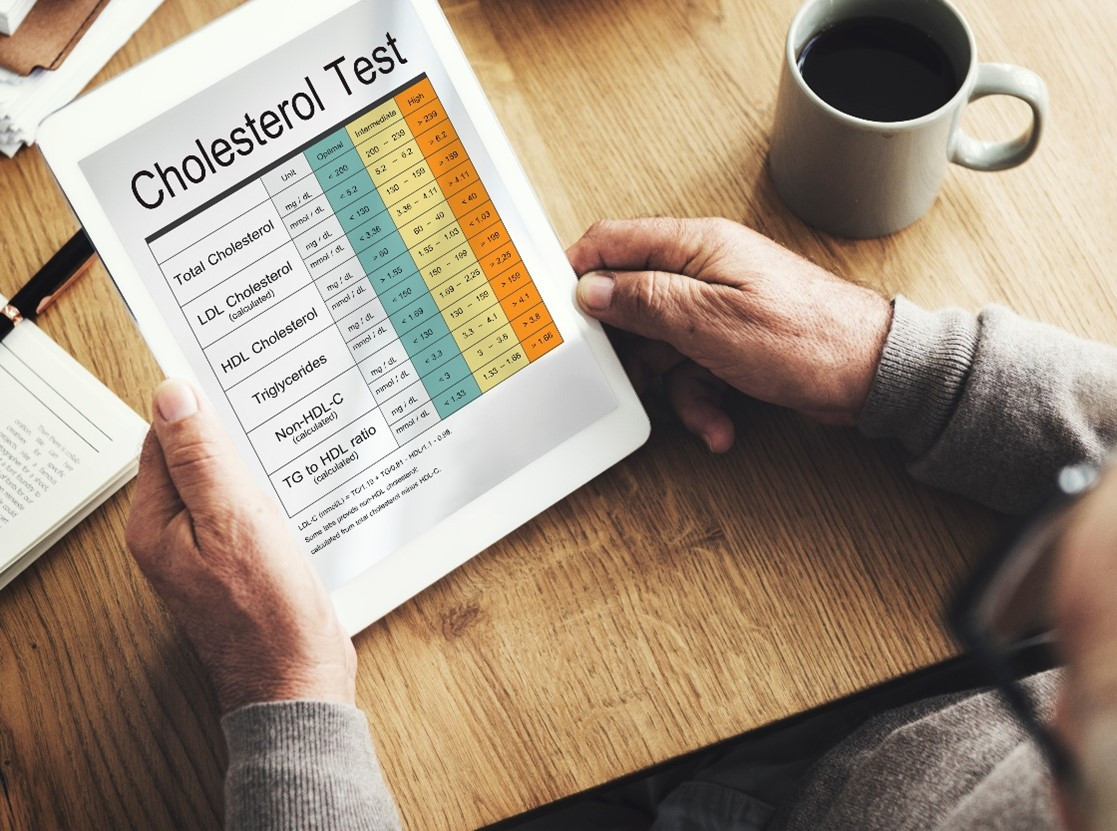
a. Konsentrasi ALT (SGPT) dan atau AST (SGOT) biasanya mengalami peningkatan ringan sampai sedang, mencapai 1-4 kali dari batas atas nilai normal dengan rasio AST/ALT kurang dari 1. Rasio tersebut khas bagi NAFLD walaupun hal tersebut tergantung pada keparahan penyakit; di mana sebaliknya, yaitu rasio AST/ALT lebih dari 1 berhubungan dengan fibrosis dan progresi penyakit. Namun, perlemakan hati alkoholik yang tidak terlalu parah juga memiliki rasio AST/ALT kurang dari 1, oleh karena itu hal ini dapat berguna untuk membedakan perlemakan hati alkoholik dari non-alkoholik, namun diperlukan interpretasi yang hati-hati.
b. Gamma-glutamiltranspeptidase (GGT) hampir selalu meningkat, Alkalin Phospatase (AP) bisa meningkat beragam sampai dengan 2 kali batas normal atas. Hasil tes fungsi hati seperti albumin, bilirubin, dan waktu prothrombin biasanya normal, kecuali bila terdapat sirosis dan gagal hati. Sejumlah penelitian juga telah dilakukan dalam menemukan prediktor non-invasif dalam mendiagnosis fibrosis lanjut dan sirosis pada pasien NAFLD, antara lain FibroTest, Hepascore, dan APRI (AST-to-platelet Ratio Index).
2. Pencitraan
Pencitraan abdomen sering dilakukan dalam mengonfirmasi kecurigaan NAFLD. Keberadaan lemak pada hati dapat diketahui melalui berbagai pencitraan non-invasif. Pada praktik sehari-hari, steatosis sering dideteksi melalui ultrasonografi (USG), computerised axial tomography (CT), dan magnetic resonance imaging (MRI), bila jumlah lemak telah melebihi 25-30% berat hati.
3. Biopsi Hati
Biopsi hati merupakan satu-satunya cara dalam mendiagnosis keberadaan serta derajat keparahan spektrum histologis NAFLD. Biopsi hati diperlukan apabila teknik pencitraan tidak dapat mendiagnosis, dan untuk mengonfirmasi keberadaan NASH, fibrosis, dan/atau sirosis.
Tatalaksana dan Perawatan
Strategi tatalaksana NAFLD dimulai dari modifikasi gaya hidup, yang dapat dilanjutkan dengan terapi menarget komponen sindrom metabolik, farmakoterapi, hingga tatalaksana sirosis. Secara umum, tujuan tatalaksana NAFLD/NASH berupa peningkatan kelangsungan hidup pasien (survival) dan menurunkan angka komplikasi. Selain itu, tujuan jangka pendek untuk resolusi kondisi NASH, memperbaiki atau mencegah perburukan fibrosis, dan memperbaiki kondisi sindrom metabolik atau risiko lain.
a. Modifikasi gaya hidup
Perubahan gaya hidup, termasuk penurunan berat badan, perubahan diet, dan latihan fisik menghasilkan perbaikan pada pasien NAFLD. Penurunan berat badan sebesar =7% total berat badan awal menurunkan steatosis, inflamasi, ballooning, serta skor aktivitas NAFLD (NAS).
b. Farmakoterapi
a. Peningkat Sensitivitas Insulin (Insulin-Sensitizing Drugs). Resistensi insulin memainkan peran sentral pada patogenesis NAFLD. Pengamatan pada tikus dengan resistensi insulin dan perlemakan hati, setelah diberi metformin atau thiazolidinediones mengalami perbaikan untuk kedua keadaan tersebut.
b. Antioksidan. Stres oksidatif merupakan mekanisme kunci dari cedera sel hati dan progresi NAFLD. Vitamin E merupakan salah satu antioksidan yang sering diteliti sebagai alternatif pengobatan NAFLD.
c. Obat Penurun Lipid (Lipid Lowering Drugs). Mengingat NAFLD sebagai kelainan homeostasis lemak hati, pemberian obat penurun kadar lemak juga menjadi salah satu pertimbangan.
d. Ursodeoxycholic Acid (UDCA) dan Asam Lemak Omega-3. Beberapa studi berusaha meneliti penggunaan UDCA (dosis konvensional dan dosis tinggi). Penggunaan asam lemak omega-3 disetujui di Amerika Serikat dalam penanganan hipertrigliserida dan sedang diteliti dalam penggunaannya dalam mengobati NAFLD.
Referensi:
1. Setiawan SI, Kurniawan J. Pilihan tatalaksana penyakit perlemakan hati non-alkohol (non-alcoholic fatty liver disease/ NAFLD). Cermin Dunia Kedokteran 2021;293:48(3):173-5.
2. Non-alcoholic fatty liver disease (NAFLD) [Internet]. [cited 2021 Aug 26]. Available from: http://eprints.undip.ac.id/44142/3/BAB_2.pdf