


Urolithiasis (Batu Saluran Kemih)
Pendahuluan dan Fakta
Batu ginjal yang terbentuk dalam ginjal disebut dengan nefrolithiasis. Urolithiasis adalah suatu kondisi yang terjadi ketika batu ginjal yang terbentuk dalam ginjal keluar dan berpindah ke bagian lain sistem saluran kemih, yang meliputi ureter, kandung kemih, dan uretra. Berikut ini adalah istilah penyakit batu bedasarkan letak batu antara lain:
· Ureterolithiasis disebut batu pada ureter
· Vesikolithiasis disebut sebagai batu pada kandung kemih/ batu buli
· Uretrolithiasis disebut sebagai batu pada uretra
Prevalensi urolithiasis meningkat dan terutama memengaruhi populasi usia produktif. Laki-laki (10,6%) lebih sering mengalami gejala dibandingkan dengan perempuan (7,1%). Seseorang yang mengalami obesitas dan kelebihan berat badan lebih rentan terhadap urolithiasis dibandingkan individu dengan berat badan normal, dan penelitian menunjukkan bahwa obesitas merupakan penyeimbang dalam pembentukan batu ginjal ketika membandingkan antara laki-laki dan perempuan.
Patofisiologi
Banyak faktor yang menyebabkan berkurangnya aliran urin dan menyebabkan obstruksi, salah satunya adalah statis urin dan menurunnya volume urin akibat dehidrasi serta ketidakadekuatan intake cairan, hal ini dapat meningkatkan risiko terjadinya urolithiasis. Rendahnya aliran urin adalah gejala abnormal yang umum terjadi (Colella, et al., 2005), selain itu, berbagai kondisi pemicu terjadinya urolithiasis seperti komposisi batu yang beragam menjadi faktor utama bekal identifikasi penyebab urolithiasis.
Faktor Risiko
Pada umumnya urolithiasis terjadi akibat berbagai sebab yang disebut faktor risiko. Terapi dan perubahan gaya hidup merupakan intervensi yang dapat mengubah faktor risiko, namun ada juga faktor risiko yang tidak dapat diubah.
1) Jenis Kelamin. Pasien dengan urolithiasis umumnya terjadi pada laki-laki 70-81% dibandingkan dengan perempuan 47-60%, salah satu penyebabnya adalah adanya peningkatan kadar hormon testosteron dan penurunan kadar hormon estrogen pada laki-laki dalam pembentukan batu
2) Umur. Urolithiasis banyak terjadi pada usia dewasa dibanding usia tua, namun bila dibandingkan dengan usia anak-anak, maka usia tua lebih sering terjadi
3) Riwayat Keluarga. Pasien yang memiliki riwayat keluarga dengan urolithiasis ada kemungkinan membantu dalam proses pembentukan batu saluran kemih pada pasien (25%) hal ini mungkin disebabkan karena adanya peningkatan produksi jumlah mucoprotein pada ginjal atau kandung kemih yang dapat membentuk kristal dan membentuk menjadi batu atau calculi
4) Kebiasaan diet dan obesitas. Intake makanan yang tinggi sodium, oksalat yang dapat ditemukan pada teh, kopi instan, minuman soft drink, kokoa, arbei, jeruk sitrun, dan sayuran berwarna hijau terutama bayam dapat menjadi penyebab terjadinya batu
5) Faktor lingkungan. Faktor yang berhubungan dengan lingkungan seperti letak geografis dan iklim. Beberapa daerah menunjukkan angka kejadian urolithiasis lebih tinggi daripada daerah lain
6) Pekerjaan. Pekerjaan yang menuntut untuk bekerja di lingkungan yang bersuhu tinggi serta intake cairan yang dibatasi atau terbatas dapat memacu kehilangan banyak cairan dan merupakan resiko terbesar dalam proses pembentukan batu karena adanya penurunan jumlah volume urin
7) Cairan. Asupan cairan dikatakan kurang apabila < 1 liter/ hari, kurangnya intake cairan inilah yang menjadi penyebab utama terjadinya urolithiasis khususnya nefrolithiasis karena hal ini dapat menyebabkan berkurangnya aliran urin/ volume urin (Domingos & Serra, 2011).
Gejala Klinis dan Komplikasi
Urolithiasis dapat menimbulkan berbagai gejala tergantung pada letak batu, tingkat infeksi, dan ada tidaknya obstruksi saluran kemih.
Beberapa gambaran klinis yang dapat muncul pada pasien urolithiasis:
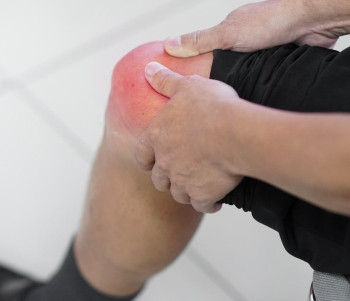
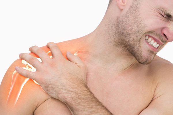
1) Nyeri pada ginjal dapat menimbulkan dua jenis nyeri yaitu nyeri kolik dan non kolik. Nyeri kolik terjadi karena adanya stagnansi batu pada saluran kemih sehingga terjadi resistensi dan iritabilitas pada jaringan sekitar.
2) Gangguan miksi. Adanya obstruksi pada saluran kemih, maka aliran urin (urine flow) mengalami penurunan sehingga sulit sekali untuk miksi secara spontan.
3) Hematuria. Batu yang terperangkap di dalam ureter (kolik ureter) sering mengalami desakan berkemih, tetapi hanya sedikit urin yang keluar. Keadaan ini akan menimbulkan gesekan yang disebabkan oleh batu sehingga urin yang dikeluarkan bercampur dengan darah (hematuria).
4) Mual dan muntah. Kondisi ini merupakan efek samping dari kondisi ketidaknyamanan pada pasien karena nyeri yang sangat hebat sehingga pasien mengalami stress yang tinggi dan memacu sekresi HCl pada lambung.
5) Demam, terjadi karena adanya kuman yang menyebar ke tempat lain.
6) Distensi vesika urinaria. Akumulasi urin yang tinggi melebihi kemampuan vesika urinaria akan menyebabkan vasodilatasi maksimal pada vesika. Oleh karena itu, akan teraba bendungan (distensi) pada waktu dilakukan palpasi pada regio vesika.
Diagnosis
Menurut Brunner & Suddart, (2015) dan Purnomo, (2012) diagnosis urolithiasis dapat ditegakkan melalui beberapa pemeriksaan seperti:
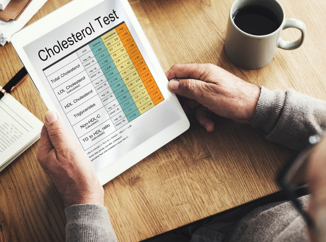
1) Kimiawi darah dan pemeriksaan urin 24 jam untuk mengukur kadar kalsium, asam urat, kreatinin, natrium, pH dan volume total (Portis & Sundaram, 2001).
2) Analisis kimia dilakukan untuk menentukan komposisi batu.
3) Kultur urin dilakukan untuk mengidentifikasi adanya bakteri dalam urin (bacteriuria) (Portis & Sundaram, 2001).
4) Foto polos abdomen
5) Intra Vena Pielografi (IVP). IVP merupakan prosedur standar dalam menggambarkan adanya batu pada saluran kemih.
6) Ultrasonografi (USG). USG sangat terbatas dalam mendiagnosa adanya batu dan merupakan manajemen pada kasus urolithiasis.
Tata Laksana
Tujuan dalam panatalaksanaan medis pada urolithiasis adalah untuk menyingkirkan batu, menentukan jenis batu, mencegah penghancuran nefron, mengontrol infeksi, dan mengatasi obstruksi yang mungkin terjadi. Tata laksana penyakit ini berdasarkan gejala akut pasien dan mencakup terapi medis konservatif dan intervensi bedah. Mengendalikan rasa nyeri pada pasien sangatlah penting. Pemberian obat antiinflamasi oral atau intravena digunakan sebagai pengobatan lini pertama pada nyeri. Mual dan muntah harus diobati dengan pemberian obat antiemetik intravena, seperti ondansetron, metoclopramide, promethazine, dan lainnya. Penggunaan obat alpha-blocker seperti doxazosin dan tamsulosin dapat membantu keluarnya batu yang ukurannya lebih besar (5-10 mm), namun obat ini belum terbukti bermanfaat untuk mengeluarkan batu yang berukuran lebih kecil.
Pasien yang datang dengan batu besar, atau jika gejalanya konsisten dengan gagal ginjal akut, oliguria/anuria, kriteria SIRS, infeksi terkait, atau riwayat penyakit ginjal, memerlukan intervensi spesialis urologi segera. Nyeri atau muntah yang tidak dapat diatasi, ketidakmampuan untuk mendapatkan asupan oral, kehamilan, atau pasien anak, maka memerlukan rawat inap untuk observasi lebih lanjut.
Intervensi lebih lanjut harus segera didiskusikan dengan spesialis urologi, dan rencana perawatan yang tepat harus dibuat sesuai dengan faktor risiko pasien, riwayat kesehatan, gejala akut, serta kenyamanan dan saran dari spesialis urologi. Ada berbagai metode intervensi urologi akut, termasuk extracorporeal shockwave lithotripsy (ESWL), flexible ureteroscopy (URS), dan percutaneous nephrolithotomy (PCNL).
Referensi:
- Urolithiasis [Internet]. Available from: http://repository.umy.ac.id/bitstream/handle/123456789/7842/6.%20BAB%20II.pdf?sequ
- Thakore P, Liang TH. Urolithiasis. National Library of Medicine [Internet]. 2023. Available from: https://www.ncbi.nlm.nih.gov/books/NBK559101/