


Hernia Nukleus Pulposus (HNP)
Pendahuluan dan Fakta
Herniasi nukleus pulposus adalah penyebab paling umum nyeri skiatik dan salah satu indikasi paling umum untuk operasi tulang belakang di seluruh dunia. Kondisi ini muncul karena perpindahan nukleus pulposus melampaui ruang diskus intervertebralis. Anatomi diskus sendiri terdiri dari dua struktur utama, nukleus pulposus dan annulus fibrosus. Nukleus pulposus terdiri dari air, kolagen tipe II, sel mirip kondrosit, dan proteoglikan. Komposisi unik ini memungkinkan nukleus pulposus menjadi elastis, fleksibel di bawah tegangan, dan menyerap penyempitan atau kompresi.
Komposisi annulus fibrosus terutama berupa lapisan serat kolagen tipe I, membentuk jaringan fibrosa dengan disposisi heliks yang mengelilingi nukleus pulposus, struktur ini lebih padat di bagian anterior dan melekat pada vertebra melalui serabut Sharpey atau Sharpey fibers.
Perkiraan prevalensi herniasi diskus adalah sekitar 1%-3%. Insiden tertinggi yang diamati adalah antara usia 30 hingga 50 tahun, dan lebih sering terjadi pada pria dibandingkan wanita.
Patofisiologi
Herniasi diskus merupakan akibat dari perubahan degeneratif pada annulus; perubahan tersebut adalah modifikasi adaptif terkait usia pada struktur diskus yang meliputi pengeringan, retakan, penyempitan diskus, degenerasi musinosa, gas intradiscal (vakum), osteofit, perubahan inflamasi, dan sklerosis subkondral. Fisura annulus merupakan predisposisi adanya kelemahan, yang memungkinkan material diskus menonjol atau bermigrasi ke luar batas annulus.
Etiologi
Herniasi diskus dan degenerasi diskus merupakan istilah yang berikatan karena herniasi nukleus pulposus merupakan evolusi dari diskus degeneratif. Degenerasi diskus biasanya dikaitkan dengan hilangnya proteoglikan. Berbagai faktor memengaruhi proses degeneratif seperti genetik, mekanik, dan perilaku.
Diskus intervertebralis adalah struktur yang memberikan fleksibilitas dan menyebarkan beban tubuh melalui tulang belakang. Beban mekanik penting dalam menjaga kesehatan diskus dengan menghasilkan sinyal ke sel yang mengatur homeostasis matriks yang tepat. Akan tetapi, paparan hipo atau hiper-loading dalam waktu lama berkorelasi dengan induksi degenerasi diskus.
Faktor Risiko
Beberapa faktor yang dapat meningkatkan risiko terjadinya hernia nukleus pulposus adalah:
- Berat badan berlebih
- Pekerjaan yang menuntut secara fisik
- Terlalu sering menyetir
- Kehidupan yang sedenter
- Genetik
- Merokok
Gejala Klinis dan Komplikasi
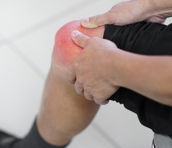
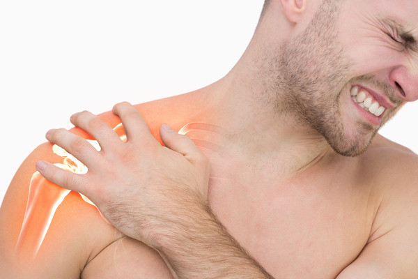
Manifestasi klinis utama yang muncul adalah rasa nyeri di punggung bawah disertai otot-otot sekitar lesi dan nyeri tekan. HNP terbagi atas HNP sentral dan lateral. HNP sentral akan menimbulkan paraparesis flasid, parestesia dan retensi urine. Sedangkan HNP lateral bermanifestasi pada rasa nyeri dan nyeri tekan yang terletak pada punggung bawah, di tengah-tengah area bokong dan betis, belakang tumit dan telapak kaki. Kekuatan ekstensi jari kelima kaki berkurang dan reflex achiller negative.
Pada HNP lateral L5-S1 rasa nyeri dan nyeri tekan didapatkan di punggung bawah, bagian lateral pantat, tungkai bawah bagian lateral, dan di dorsum pedis. Kelemahan m. gastrocnemius (plantar fleksi pergelangan kaki), m. ekstensor halusis longus (ekstensi ibu jari kaki). Gangguan reflex Achilles, defisit sensorik pada malleolus lateralis dan bagian lateral pedis (Setyanegara dkk, 2014).
Diagnosis
Pemeriksaan Neurologis, untuk memastikan bahwa nyeri yang timbul termasuk dalam gangguan saraf. Meliputi pemeriksaan sensoris, motorik, reflex.
a. Pemeriksaan sensoris, pada pemeriksaan sensoris ini apakah ada gangguan sensoris, dengan mengetahui dermatom mana yang terkena akan dapat diketahui radiks mana yang terganggu.
b. Pemeriksaan motorik, apakah ada tanda paresis, atropi otot.
c. Pemeriksaan reflex, bila ada penurunan atau refleks tendon menghilang, misal APR menurun atau menghilang berarti menunjukkan segmen S1 terganggu.
Adapun tes yang dapat dilakukan untuk diagnosis hernia nucleus pulposus (HNP) adalah:
a. Pemeriksaan Range of Movement (ROM)
Pemeriksaan ini dapat dilakukan secara aktif oleh penderita sendiri ataupun secara pasif oleh pemeriksa. Pemeriksaan ROM ini memperkirakan derajat nyeri, function laesa, atau untuk memeriksa ada/ tidaknya penyebaran rasa nyeri.
b. Straight Leg Raise (SLR) Test.
Tes untuk mengetaui adanya jebakan nervus ischiadicus. Pasien tidur dalam posisi supinasi dan pemeriksa memfleksikan panggul secara pasif, dengan lutut dari tungkai terekstensi maksimal. Tes ini positif bila timbul rasa nyeri pada saat mengangkat kaki dengan lurus, menandakan ada kompresi dari akar saraf lumbar.
c. Lasegue Menyilang.
Caranya sama dengan percobaan lasegue, tetapi disini secara otomatis timbul pula rasa nyeri ditungkai yang tidak diangkat. Hal ini menunjukkan bahwa radiks yang kontralateral juga turut tersangkut.
d. Tanda Kerning
Pada pemeriksaan ini penderita yang sedang berbaring difleksikan pahanya pada persendian panggung sampai membuat sudut 90 derajat. Selain itu tungkai bawah diekstensikan pada persendian lutut. Biasanya kita dapat melakukan ekstensi ini sampai sudut 135 derajat, antara tungkai bawah dan tungkai atas, bila terdapat tahanan dan rasa nyeri sebelum tercapai sudut ini, maka dikatakan tanda kerning positif.
e. Ankle Jerk Reflex
Dilakukan pengetukan pada tendon Achilles. Jika tidak terjadi dorsofleksi pada kaki, hal ini mengindikasikan adanya jebakan nervus di tingkat kolumna vertebra L5-S1.
f. Knee-Jerk Reflex
Dilakukan pengetukan pada tendon lutut. Jika tidak terjadi ekstensi pada lutut, hal ini mengindikasikan adanya jebakan nervus di tingkat kolumna vertebra L2-L3-L4.
Diagnosis Penunjang
X-ray adalah pemeriksaan awal ketika ada kecurigaan kuat terhadap penyebab spesifik nyeri tulang servikal atau punggung (patah tulang, infeksi, tumor) atau jika ada tanda bahaya (demam, usia lebih dari 50 tahun, trauma baru-baru ini, nyeri di malam atau istirahat, penurunan berat badan tanpa sebab yang jelas, defisit motorik atau sensorik progresif, riwayat kanker atau osteoporosis, kegagalan untuk membaik setelah enam minggu pengobatan konservatif). Rontgen anteroposterior dan lateral berguna untuk menilai fraktur, deformitas tulang, penurunan tinggi intervertebralis, osteofit, spondilolistesis, dan osteoartritis sendi.
MRI adalah pencitraan diagnostik yang direkomendasikan dalam kasus defisit neurologis yang parah atau progresif, kecurigaan terhadap kondisi yang mendasari seperti infeksi, patah tulang, sindrom cauda equina, kompresi sumsum tulang belakang. Dalam kasus radikulopati, sebagian besar kasus membaik dengan pengobatan konservatif dan MRI diindikasikan pada kasus dengan nyeri yang signifikan atau defisit neurologis. CT myelogram adalah pilihan pencitraan pada pasien dengan kontraindikasi terhadap MRI.
CT scan biasanya tidak diminta pada herniasi nukleus pulposus. Namun, hal ini dapat membantu dalam beberapa kasus ketika ada kecurigaan adanya kalsifikasi herniasi diskus (herniasi diskus toraks memiliki tingkat kalsifikasi sebesar 30 hingga 70%) yang lebih sulit terutama bila pembedahan menjadi pertimbangan.
Tata Laksana dan Perawatan
Terapi Non-Farmakologis, yaitu:
a. Kompres hangat/dingin
Kompres hangat/dingin ini merupakan modalitas yang mudah dilakukan. Untuk mengurangi spasme otot dan inflamasi. Beberapa pasien merasakan nyeri hilang pada pengkompresan hangat, sedangkan yang lain pada pengkompresan dingin.
b. Iontophoresis
Merupakan metode pemberian steroid melalui kulit. Steroid tersebut menimbulkan efek anti inflamasi pada daerah yang menyebabkan nyeri. Modalitas ini terutama efektif dalam mengurangi serangan nyeri akut.
c. Unit TENS (Transcutaneous Electrical Nerve Stimulator)
Sebuah unit transcutaneous electrical nerve stimulator (TENS) menggunakan stimulasi listrik untuk mengurangi sensasi nyeri punggung bawah dengan mengganggu impuls nyeri yang dikirimkan ke otak
d. Ultrasound, merupakan suatu bentuk penghangatan di lapisan dalam dengan menggunakan gelombang suara pada kulit yang menembus sampai jaringan lunak dibawahnya. Ultrasound terutama berguna dalam menghilangkan serangan nyeri akut dan dapat mendorong terjadinya penyembuhan jaringan.
Latihan dan modifikasi gaya hidup
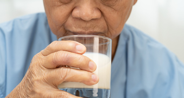
Berat badan yang berlebihan harus diturunkan karena akan memperberat tekanan ke punggung bawah. Program diet dan latihan penting untuk mengurangi NPB pada pasein yang mempunyai berat badan berlebihan. Direkomendasikan untuk memulai latihan ringan tanpa stres secepat mungkin. Endurance exercisi latihan aerobik yang memberi stres minimal pada punggung seperti jalan, naik sepeda atau berenang dimulai pada minggu kedua setelah awaitan NPB. Conditional execise yang bertujuan memperkuat otot punggung dimulai sesudah dua minggu karena bila dimulai pada awal mungkin akan memperberat keluhan pasien. Latihan memperkuat otot punggung dengan memakai alat tidak terbukti lebih efektif daripada latihan tanpa alat.
Terapi Farmakologis
a. Analgetik dan NSAID (Non Steroid Anti Inflamation Drug) obat ini diberikan dengan tujuan untuk mengurangi nyeri dan inflamasi sehingga mempercepat kesembuhan. Contoh analgetik : paracetamol, Aspirin Tramadol. NSAID : Ibuprofen, Natrium diklofenak, Etodolak, Selekoksib.
b. Obat pelemas otot (muscle relaxant) bermanfaat bila penyebab NPB adalah spasme otot. Efek terapinya tidak sekuat NSAID, seringkali di kombinasi dengan NSAID. Sekitar 30% memberikan efek samping mengantuk.
Contoh Tinazidin, Esperidone dan Carisoprodol.
c. Opioid. Obat ini terbukti tidak lebih efektif daripada analgetik biasa yang jauh lebih aman. Pemakaian jangka panjang bisa menimbulkan toleransi dan ketergantungan obat.
d. Kortikosteroid oral Pemakaian kortikosteroid oral masih kontroversi. Dipakai pada kasus HNP yang berat dan mengurangi inflamasi jaringan.
e. Anelgetik ajuvan, terutama dipakai pada HNP kronis karena ada anggapan mekanisme nyeri pada HNP sesuai dengan neuropatik. Contohnya : amitriptilin, Karbamasepin, Gabapentin.
f. Suntikan pada titik picu
Cara pengobatan ini dengan memberikan suntikan campuran anastesi lokal dan kortikosteroid ke dalam jaringan lunak/otot pada titik picu disekitar tulang punggung. Cara ini masih kontroversi. Obat yang dipakai antara lain lidokain, lignokain, deksametason, metilprednisolon dan triamsinolon.
Terapi Operatif
Dilakukan pada pasien dilakukan jika:
a. Pasien mengalami HNP grade 3 atau 4.
b. Tidak ada perbaikan lebih baik, masih ada gejala nyeri yang tersisa, atau ada gangguan fungsional setelah terapi konservatif diberikan selama 6 sampai 12 minggu.
c. Terjadinya rekurensi yang sering dari gejala yang dialami pasien menyebabkan keterbatasan fungsional kepada pasien, meskipun terapi konservatif yang diberikan tiap terjadinya rekurensi dapat menurunkan gejala dan memperbaiki fungsi dari pasien.
d. Terapi yang diberikan kurang terarah dan berjalan dalam waktu lama.
Pilihan terapi operatif yang dapat diberikan adalah:
a. Distectomy Pengambilan sebagian diskus intervertabralis.
b. Percutaneous distectomy. Pengambilan sebagian diskus intervertabralis dengan menggunakan jarum secara aspirasi.
c. Laminotomy/laminectomy/foraminotomy/facetectomy
Melakukan dekompresi neuronal dengan mengambil beberapa bagian dari vertebra baik parsial maupun total.
d. Spinal fusion dan sacroiliac joint fusion: Penggunaan graft pada vertebra sehingga terbentuk koneksi yang rigid diantara vertebra sehingga terjadi stabilitas
Referensi:
- De Cicco FL, Willhuber GO. Nucleus pulposus herniation. National Library of Medicine [Internet]. 2023. Available from: https://www.ncbi.nlm.nih.gov/books/NBK542307/#article-22825.s2
- Mayo Clinic. Herniated disk [Internet]. 2023. Available from: https://www.mayoclinic.org/diseases-conditions/herniated-disk/symptoms-causes/syc-20354095
- HNP [Internet]. Available from: https://sinta.unud.ac.id/uploads/dokumen_dir/464eb57bfb86f13cb48f856a325ff96a.pdf
- Hernia Nucnleus Pulposus [Internet]. Available from: https://med.unhas.ac.id/kedokteran/wp-content/uploads/2016/09/Bahan-Ajar-4_Hernia-Nucleus- Pulposus.pdf